Mechanics of Breathing

Breathing is due to changes of pressure within the thorax in comparison with the outside.

The diaphragm is the most important muscle for inspiration.

It is attached to the lower portion of the rib cage and—when contracted—pushes the contents of the abdomen downward, which increases the vertical dimension of the thorax.

Active downward movement or contraction of the diaphragm generates an increase in negative pleural pressure that causes the lungs to expand and fill with air during inspiration.

The contraction of the diaphragm also pulls up on the naturally downsloping rib cage that is hinged to the vertebrae in a bucket-handle fashion. Lifting the individual bucket handles acts to increase the anteroposterior dimension of the thorax . The increased volume of the thorax creates a negative intrathoracic pressure that draws air through the airways and into the alveoli.

++++++++++++++++


During expiration, this negative pressure decreases and the air is passively expelled. During inspiration, air is filtered, warmed, and humidified as it travels through the upper and lower airways. Cilia, hairlike structures within the passageways, help to move air toward the alveoli. Cilia also help to move mucus and debris out of the pulmonary system, keeping the lower airways from being contaminated. The sterility of the lower airway is achieved with the help of mucus-secreting goblet cells. Mucus traps debris and keeps the airway moist. Gas exchange occurs in the alveoli and pulmonary capillaries. Oxygen and carbon dioxide are exchanged at this cellular level.

+++++++++++++++

At the same time, venous blood returning from the various body tissues is pumped into the lungs by the right ventricle of the heart. This mixed venous blood has a high carbon dioxide content and a low oxygen content. In the pulmonary capillaries, carbon dioxide is exchanged for oxygen from the alveoli. The blood leaving the lungs, which now has a high oxygen content and a relatively low carbon dioxide content, is distributed to the tissues of the body by the left side of the heart. During expiration, gas with a high concentration of carbon dioxide is expelled from the body.

++++++++++++++++++++


The diaphragm is innervated by the phrenic nerve that originates from the cervical levels C3–C5.

The diaphragm is unique in that it acts as both an involuntary and voluntary muscle. During sleep and rest, the diaphragm contracts involuntarily at a rate determined by respiratory centers in the medulla. This process can be overcome voluntarily by conscious breath holding, increased inspiratory excursion, or forceful exhaling. 

++


Passive upward movement or relaxation of this crucial muscle causes an increase in pleural pressure and results in an increased pressure in the airways, facilitating expiration. The diaphragm is innervated by the phrenic nerve , which originates from the C3, C4, and C5 spinal nerves. In addition to the motor fibers to the diaphragm, the phrenic nerve also contains pain fibers, which is why pain originating from the diaphragm can be referred to the shoulders.

++++++++++++++++++++++


Air moves from a region of higher pressure to one of lower pressure.

Atmospheric pressure is conventionally referred to as 0 cm H2O, so lowering alveolar pressure below atmospheric pressure is known as negative-pressure breathing. 


+++++++++++++++++++++


The lungs and the chest wall are elastic structures.

Because of the thin layer of fluid in the intrapleural space, the lungs slide easily on the chest wall, but resist being pulled away from it in the same way that two moist pieces of glass slide on each other but resist separation. The pressure in the “space” between the lungs and chest wall (intrapleural pressure) is subatmospheric 

Image not available.
Pressure in the alveoli and the pleural space relative to atmospheric pressure during inspiration and expiration.
 The dashed line indicates what the intrapleural pressure would be in the absence of airway and tissue resistance; the actual curve (solid line) is skewed to the left by the resistance. Volume of breath during inspiration/expiration is graphed for comparison.


It is also possible to cause air to flow into the lungs by raising the pressure at the nose and mouth above alveolar pressure.


This positive-pressure ventilation is generally used on patients unable to generate a sufficient pressure difference between the atmosphere and the alveoli by normal negative-pressure breathing.

Air flows out of the lungs when alveolar pressure is sufficiently greater than atmospheric pressure to overcome the resistance to airflow offered by the conducting airways.


++++++++++++++++++

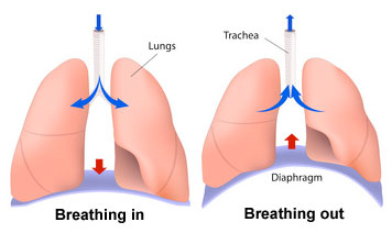
On exhalation, the diaphragm and intercostal muscles relax and return to their resting positions. This reduces the size of the thoracic cavity, thereby increasing the pressure and forcing air out of the lungs.

++++++++++++++Air is moved through the lungs by a ventilating mechanism, consisting of the thoracic cage, intercostal muscles, diaphragm, and elastic components of the lung tissue.


Mechanics of Breathing

Digital World Medical School
© 2025